What is a Breast Biopsy?
If your clinicians are concerned about a spot in your breast, they may need more information to make a diagnosis. A breast biopsy is when a clinician takes a small piece of tissue from the spot. The tissue is examined very closely under a microscope to make a diagnosis. Often the spot is not serious, and no treatment is needed. Sometimes a breast biopsy will diagnose something more serious like cancer. It can help your clinician determine what types of treatment to offer you.
How is a Biopsy done?
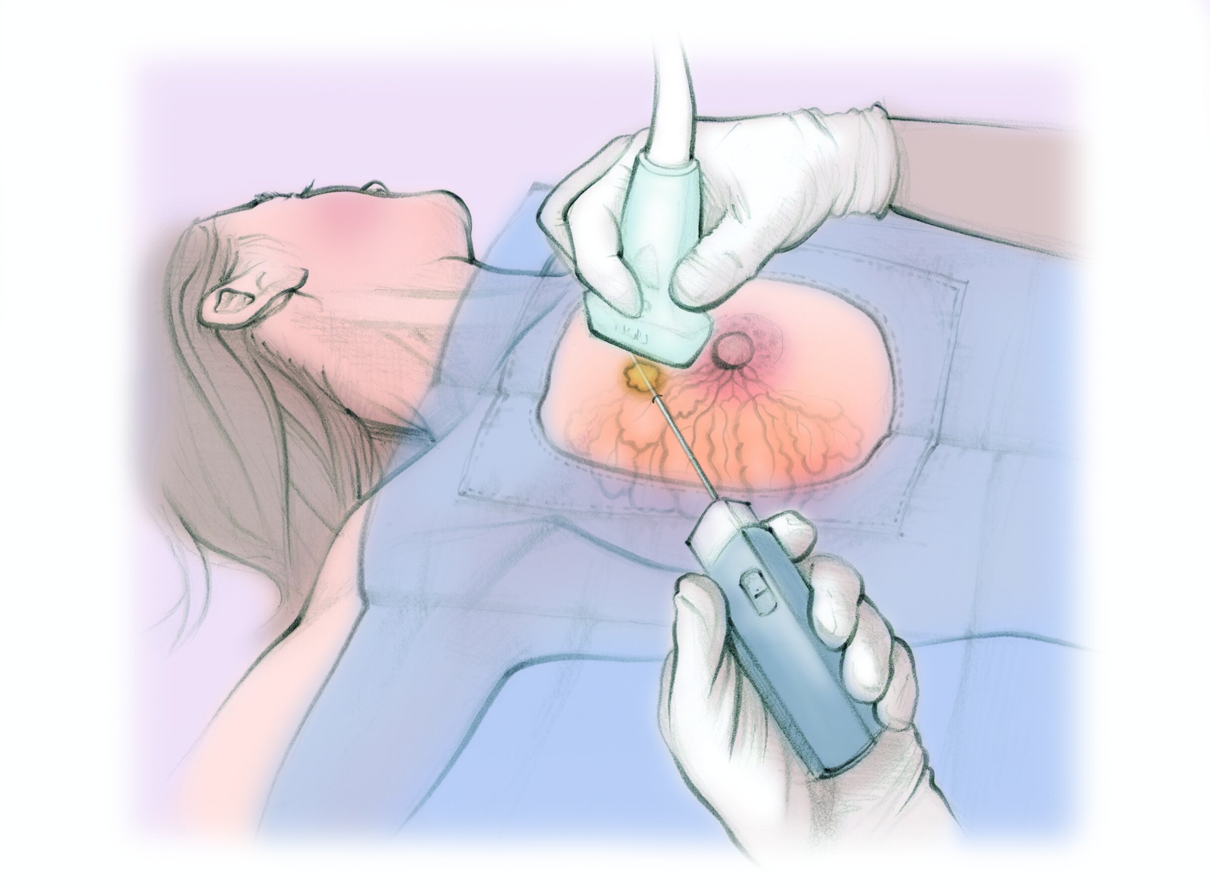
A breast biopsy is done with the help of medical imaging to show the spot inside the breast. If it is done with ultrasound, you will lay on your back. If it is done with a mammogram, you will sit in a chair or on your belly. If it is done with MRI, you will lay on your belly.
The clinician first finds the spot using imaging then numbs the breast. The clinician places a skinny needle into the spot through a pinhole in the skin. It is normal to feel pressure. The needle makes sounds as it collects samples.
After the biopsy, the clinician may leave a tiny metallic clip to mark the place where the biopsy was done. This clip will not set off metal detectors. It is OK to stay in your breast permanently if the spot does not need to be removed. After the biopsy, the clinician will cover the pinhole in your skin with a bandage. It will heal in a few days.
Breast Biopsy
1. The clinician finds the spot using imaging then numbs the breast.
2. The clinician places a skinny needle into the spot through a pinhole in the skin. The needle makes sounds as it collects samples.
3. The clinician leaves a tiny metallic clip to mark the place where the biopsy was done. Then they put a bandage over the pinhole in the skin.
What are the risks?
Breast biopsies are safe when done by a specialist.
Most commonly women will experience soreness at the biopsy site for a day or two.
Less than 1 in 100 people have bleeding or a large bruise.
Less than 1 in 1000 people have a serious infection
What are the alternatives?
Alternative 1 Not doing a biopsy. You may avoid a procedure but your clinicians may have trouble diagnosing the spot. This could prevent you from getting the right treatment, if you need it.
Alternative 2 Watching and waiting. You and your clinician may choose to watch the spot with follow-up imaging exams to see if it grows or changes. The downside is that you may delay treatment if it is something serious.
Alternative 3 Surgery to cut out the spot. This has more risks and a longer recovery. The surgeon may want a diagnosis before they operate.


