What is a port?
A port-a-catheter, or “port,” is a small device that provides long-term access to a patient’s vein for medicines or blood exchanges. Ports are placed completely under the skin of the upper chest.
The port is a metal or plastic well about the size of a quarter and is half an inch thick. The well is covered with a flexible roof that can be punctured with a special needle. It is connected to a thin plastic tube.
The special needle is used to puncture the port through the skin. Ports are usually more comfortable than getting stuck in the vein over and over. They can also be a good way to give some medicines to treat cancer or chronic diseases.
Ports have a very low risk of infection. Patients can have them for months to years. Once the port site has healed, patients can swim and bathe normally.
How is a port placed?
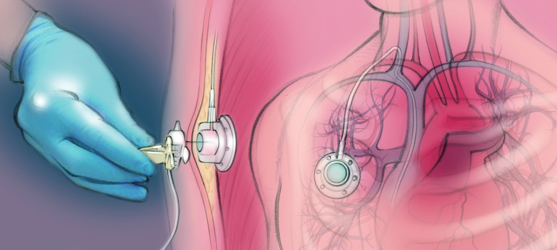
The clinician numbs the skin at the base of the neck and on the upper chest. They slide the well under the skin of the upper chest through a cut about an inch long. They tunnel the tube under the skin and into a vein in the neck. They use x-rays to make sure the tip of the tube is in a big vein near the heart. They close the cut with stitches. The port makes a small bump under the skin.
Port Placement Procedure
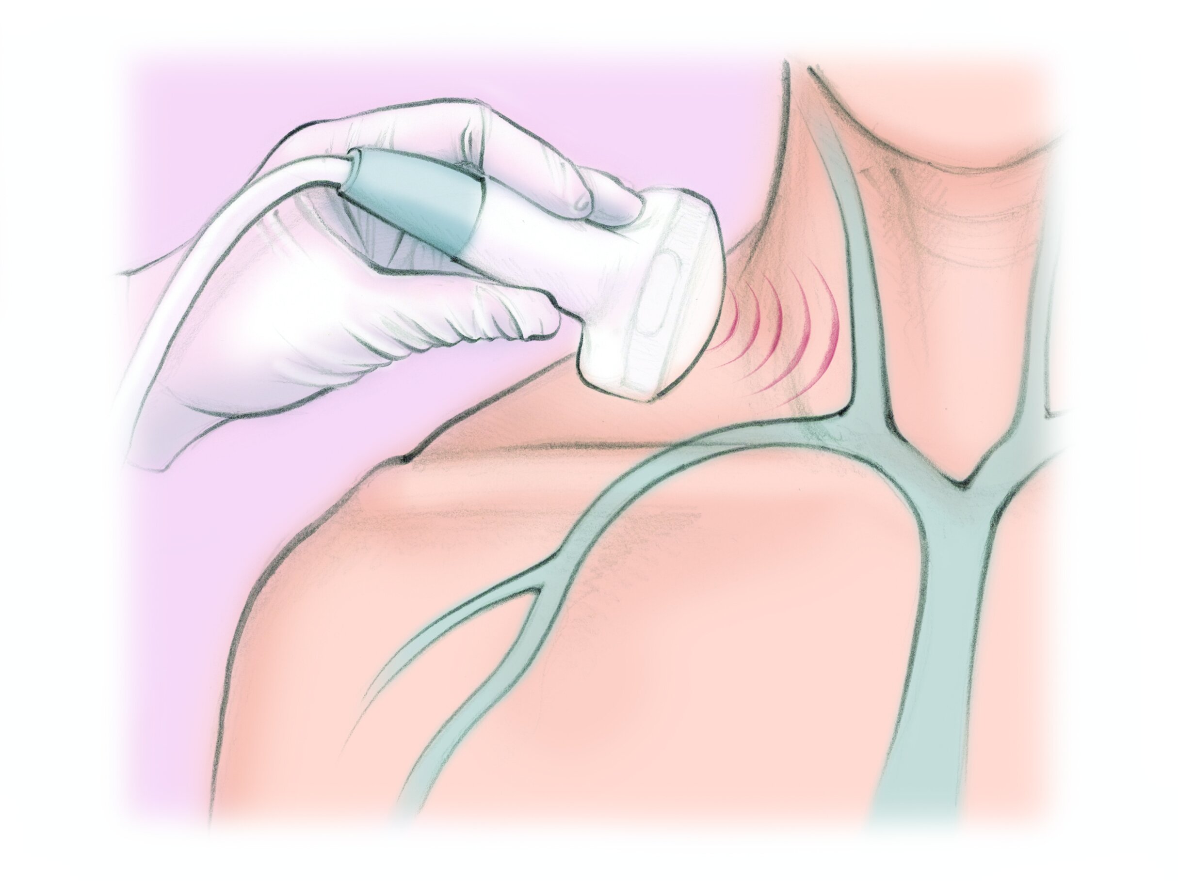
1. The clinician numbs the skin of the base of the neck and upper chest. They use ultrasound to put a small tube in a vein in the neck.
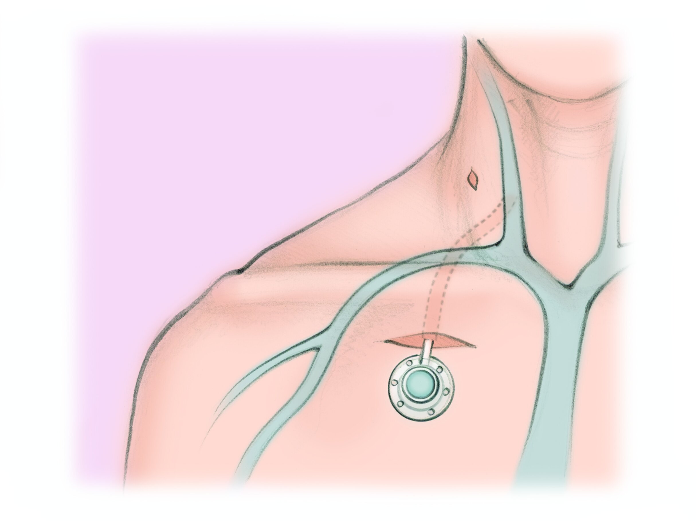
2. They tunnel the tube under the skin of the chest and connect it to the port. They slide the port into a small pocket under the skin of the the chest.
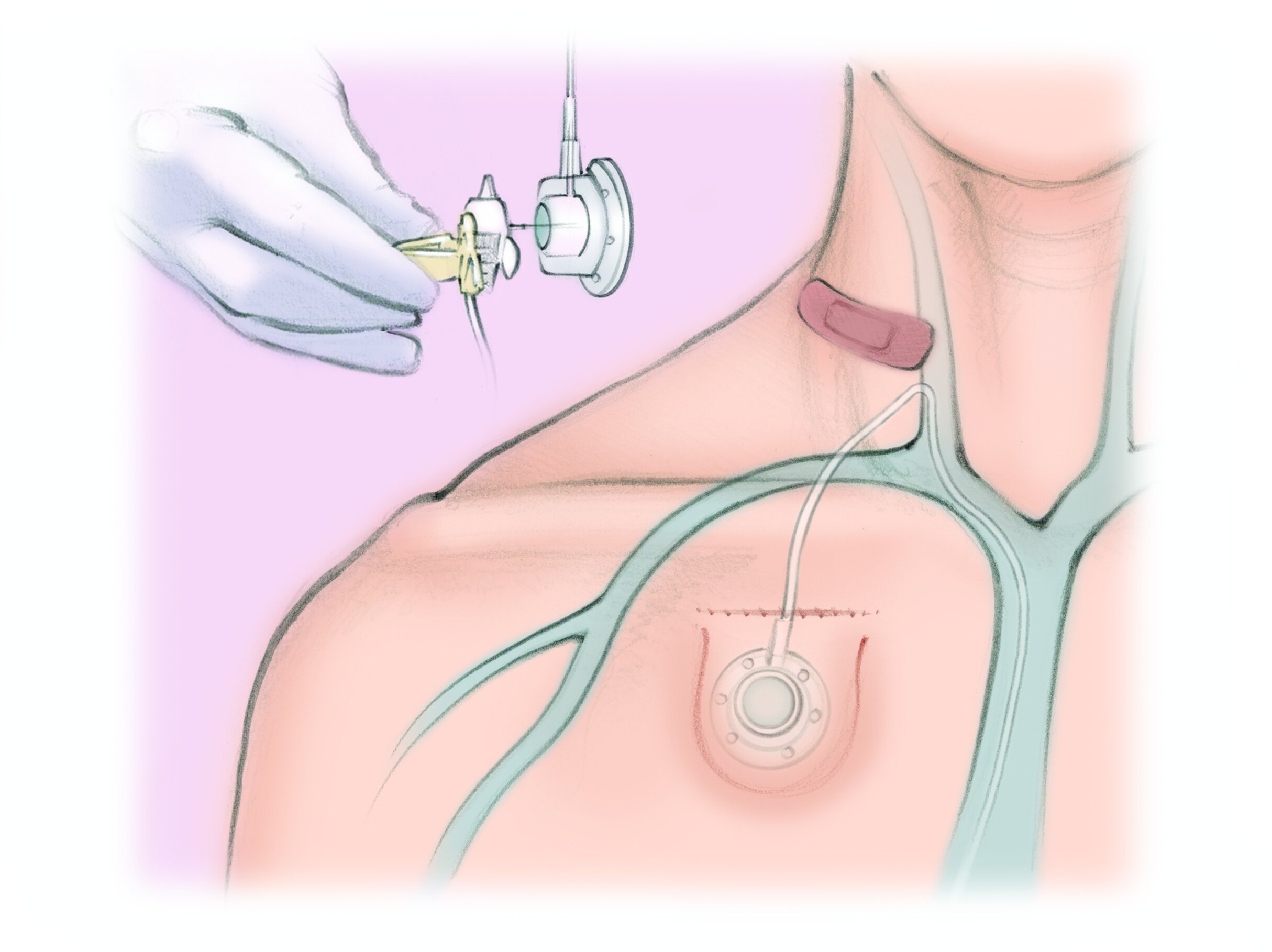
3. The clinician closes the pocket with stitches and puts a bandage over the small spot on the neck. A special needle is used to puncture the port through the skin.
What are the risks?
Port placement is generally a safe procedure when done by a specialist. Complications are rare.
Complications are rare and include infection in up to 5 in 100 patients and bleeding in 2 in 100 patients. Damage to the lung, blood vessels or nerves occur in less than 1 in 100 patients. Less than 1 in 100 patients will develop symptoms from air getting into the vein.
Over time, there is a risk of the port getting infected during use. Uncommonly, blood clots can form in or around the tube. Rarely, the port can twist in the pocket or separate from the tubing.
What are the alternatives?
Your alternatives depend on what you need vein access for and for how long.
Alternative 1 No port or other IV. The disadvantage is that you may not be able to get the treatment that you need.
Alternative 2 PICC line. A PICC is a special IV that enters the arm and ends in a big vein in the chest. PICCs require regular maintenance. They cannot get wet. The risk of infection is higher than a port. They must be removed or replaced every couple months.
Alternative 3 Regular IV. Regular IVs go into small veins. Small veins cannot handle some treatments. They need to be replaced every few days.
Frequently asked questions
What is a port?
A port (or portacath) is a small device that provides long-term access to a patient’s vein to give them medicines or draw blood.
The port sits under the skin below the collarbone or, less commonly, in the upper arm.
What are the advantages of a port over an IV or other central line?
A port allows a patient to avoid having to get stuck in the arm every time they need medicine or a blood draw. Also, some medicines are too strong for the small veins so they need to be given into the big veins near the heart.
Can stay in place for months to years
Minimal pain to use
Lower risk of infection
Patients can swim and bathe normally because the port is entirely under the skin
Who places ports?
In the past, ports were placed by a surgeon using anesthesia. Nowadays, most ports are placed by an Interventional Radiologist (IR) in a safe minimally invasive, image-guided procedure (MIIP).
How does the IR doctor place a port?
After cleaning the skin, the IR doctor numbs the skin in two spots: at the base of the neck and on the chest below the collarbone.
The IR places the port in a pocket under the skin of the chest through a short incision.
The IR tunnels the tubing under the skin to the neck then places it into the vein through a small knick in the skin.
The IR watches with X-rays while positioning the tubing near the heart.
The IR then closes the port pocket with stitches.
Is port placement a safe procedure?
This MIIP is safe for 2 main reasons:
IRs use radiology pictures to make sure the port is in the right place.
Patients only need local numbing medicine or medicine to cause drowsiness, so they do not have the risks of anesthesia.
This MIIP usually takes an hour or less. Most patients can have their port placed as an outpatient, meaning they go home the same day.
Who could benefit from a port?
Patients with cancer who need chemotherapy
Patients with chronic diseases like cystic fibrosis or other diseases requiring long-term antibiotics or blood exchanges.
Patients who need frequent blood draws but have poor vein access
For More Information:
For general information about ports, visit http://jamanetwork.com/journals/jamaoncology/fullarticle/2476249.